|
Microphotography
Light and Epifluorescence Microscopy We mounted a Nikon CoolPix 990 digital camera to a Zeiss Axioskop epifluorescence microscope for both light and fluorescence microscopy. The camera was attached to the c-mount phototube of the Axioskop by a metal sleeve. The metal sleeve fits over the standard Zeiss c-mount and possesses a thread at the upper end that fits the lense threat of the CoolPix. The camera is operated using a net adapter because the batteries' life time is too short for routine use of the camera on the microscope. Microphotographs are usually taken at the highest camera photo quality (Fine), but pictures posted here were taken at intermediate quality.
| ||||
|
These photographs demonstrate that reasonable microphotography is possible with a Nikon CoolPix digital camera. Although the quality of photographs might not reach that of professional microphotography systems, simple digital cameras present an affordable alternative for routine photodocumentation. Professional microphotography systems offered by the microscope manufacterers and specialized companies run easily at $10,000-$20,000. Digital cameras allow photodocumentation for private or hobby microscopy for less than a few hundred dollars (excl. the microscope). The here presented work was performed at the Marine Science Institute at The University of Texas at Austin. The Zeiss Axioskop microscope was funded by German Research Coucil grant DFG Jo 192/5-1. A triple tube C-mount and the Nikon Cooplix were funded by the Nancy Lee and Perry R. Bass Endowment to the University of Texas at Austin. |

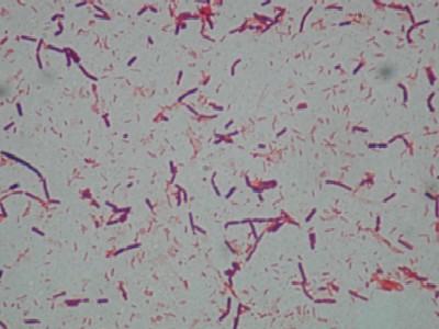 Gram-staining of bacteria isolated on general nutrient agar plates for the Gulf Stream off
the South Florida coast. Long gram-positive rods in chains appear violet, gram-negative rods
red. 100× oil immersion objective.
Gram-staining of bacteria isolated on general nutrient agar plates for the Gulf Stream off
the South Florida coast. Long gram-positive rods in chains appear violet, gram-negative rods
red. 100× oil immersion objective.